
Science & Technology · UPSC Notes
Human Body — UPSC Science & Tech Notes
Free, exam-focused notes for UPSC Prelims and Mains, with Prelims tips and Mains points for every topic. 28 topics.
Open in the interactive reader: bookmarks, progress tracking and read-aloud.
Human Digestive System: From Ingestion to Absorption
Digestion is the biochemical process of breaking down complex Biomacromolecules into simple, absorbable substances. This journey starts in the Mouth, where mechanical and chemical breakdown begins, and continues through a series of specialized organs designed to extract nutrients and energy essential for survival.
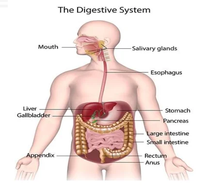
1. The Process of Digestion
Our body cannot utilize carbohydrates or proteins in their original complex form. They must be converted into their simplest units:
Carbohydrates -> Glucose (Simple Sugars)
Fats -> Fatty Acids and Glycerol
Proteins -> Amino Acids
2. The Beginning: Mouth and Buccal Cavity
The first step of the digestive process is Ingestion, taking food into the body through the mouth.
Chemical Digestion: Saliva contains Salivary Amylase, an enzyme that initiates the breakdown of starch (carbohydrates).
Protection: Saliva also contains Lysozyme, which acts as an antibacterial agent to kill harmful microbes entering with food.
The Pharynx: As food moves toward the esophagus, the Epiglottis (a cartilaginous flap) closes the glottis (windpipe opening) to prevent choking.
3. The Gastrointestinal Highway
Once swallowed, food travels through the Oesophagus via a wave-like muscular contraction called Peristalsis.
Stomach: A J-shaped bag that secretes Mucous, Hydrochloric Acid (HCl), and digestive juices. HCl kills bacteria and creates an acidic medium for protein-digesting enzymes like Pepsin.
Small Intestine: This is the site of complete digestion. It receives secretions from the Liver (Bile) and Pancreas (Pancreatic juice).
Large Intestine: Its primary role is to absorb water and salts from undigested food before the waste is expelled.
| Component | Function | Location |
|---|---|---|
| Salivary Amylase | Carbohydrate breakdown | Mouth |
| Lysozyme | Antibacterial defense | Mouth (Saliva) |
| Hydrochloric Acid (HCl) | Kills bacteria / Activates enzymes | Stomach |
| Bile | Fats emulsification | Small Intestine (from Liver) |
| Villi | Nutrient absorption | Small Intestine |
Prelims tips
- Digestion: Conversion of non-absorbable macromolecules to absorbable simple substances.
- Salivary Amylase: It specifically targets starch/carbohydrates.
- Lysozyme: A vital 'First Line of Defense' in the innate immune system within the mouth.
- Peristalsis: The involuntary constriction and relaxation of muscles in the esophagus that pushes food down.
Mains points
- Nutritional Biochemistry: The efficiency of carbohydrate, protein, and fat digestion is the foundation of metabolic health and prevents disorders like malnutrition or obesity.
- Evolutionary Adaptation: The presence of the epiglottis highlights the evolutionary solution to the shared pathway of the respiratory and digestive systems in mammals.
- Public Health: Understanding the role of lysozyme and stomach acid (HCl) explains the body's natural resistance to many common food-borne pathogens.
- Biotechnology: Modern food processing often involves 'pre-digesting' complex molecules using enzymes similar to amylase to make them easier for sensitive digestive systems to handle.
Did you know?
- An adult's small intestine is about 5 meters long! It is called 'small' only because of its narrow diameter compared to the large intestine.
- The Villi in your small intestine increase the surface area for absorption so much that if spread out, they would cover a whole tennis court.
- The Epiglottis is so efficient that you can technically swallow food even while standing on your head (though not recommended!).
Stomach and Small Intestine: Digestion and Absorption
The stomach acts as a chemical processor using Hydrochloric Acid (HCl) to break down proteins, while the Small Intestine serves as the primary site for complete digestion and nutrient absorption. The complex interaction between enzymes and structural adaptations like Villi ensures that the body successfully extracts vital energy from food.
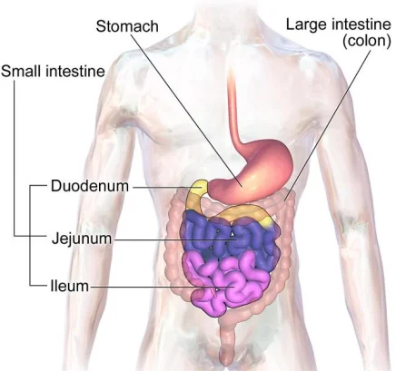
1. The Stomach: Chemical Breakdown
The stomach is a thick-walled, J-shaped bag that receives food from the food pipe. It performs three critical secretory functions:
Mucus: Protects the inner lining of the stomach from the corrosive effects of its own acid.
Hydrochloric Acid (HCl): Creates an acidic medium required for protein-digesting enzymes to work and kills many harmful bacteria that enter with food.
Digestive Juices: Specifically breaks down Proteins into simpler substances (peptones and proteoses).
2. The Small Intestine: The Absorption Hub
The small intestine is a highly coiled tube, approximately 5 meters long. It is divided into three parts: Duodenum, Jejunum, and Ileum.
Secretions: It receives bile from the liver and pancreatic juice from the pancreas to complete the digestion of fats, proteins, and carbohydrates.
Absorption via Villi: The inner walls possess thousands of finger-like outgrowths called Villi. These increase the surface area for the absorption of digested food.
Vascular Connection: Each villus has a network of thin blood vessels. Absorbed substances are transported via these vessels to different organs.
3. Assimilation and Egestion
Assimilation: The process where absorbed nutrients are used by the body to build complex substances like proteins or to produce energy (glucose breakdown with oxygen).
Final Phase: Undigested and unabsorbed food enters the large intestine for water recovery before being expelled.
| Feature | Stomach | Small Intestine |
|---|---|---|
| Primary Function | Protein digestion and disinfecting food | Complete digestion and nutrient absorption |
| Medium | Highly Acidic (due to HCl) | Alkaline (due to Bile and Pancreatic juice) |
| Length | Bag-like structure | Approximately 5 meters |
| Structural Adaptation | Rugae (folds) for expansion | Villi for surface area absorption |
| Main End Product | Chyme (partially digested food) | Simple absorbable nutrients (Glucose, etc.) |
Prelims tips
- HCl Role: It kills pathogens and activates the enzyme Pepsin.
- Small Intestine Parts: Duodenum (proximal), Jejunum (middle), Ileum (distal).
- Assimilation: Focus on the fact that this is the utilization of nutrients, not just the intake.
- Villi: Richly supplied with blood vessels to facilitate immediate transport of nutrients.
Mains points
- Metabolic Disorders: Malabsorption syndromes often stem from damage to the villi, highlighting their central role in maintaining systemic nutrition.
- Evolutionary Efficiency: The extensive length and coiled nature of the small intestine allow for maximum time and surface area to extract energy from a diverse range of diets.
- Public Health: Understanding the acidic barrier of the stomach (HCl) is fundamental in explaining why some pathogens (like Cholera) require a large dose to cause infection.
- Biochemical Coordination: The duodenum acts as a critical junction where acidic chyme from the stomach is neutralized by alkaline secretions, protecting the intestinal lining.
Did you know?
- Despite its name, the Small Intestine is the longest part of the digestive tract; it's called 'small' only because it is narrower than the large intestine.
- Your stomach produces a new layer of mucus every two weeks to prevent itself from digesting its own tissues!
- The Villi are so numerous that the total internal surface area of the small intestine is roughly the size of a studio apartment.
Large Intestine: Water Recovery and Waste Management
The Large Intestine is the final major section of the gastrointestinal tract. While the small intestine focuses on nutrient extraction, the large intestine specializes in reabsorbing water and essential salts from undigested food matter, transforming liquid waste into solid feces for Egestion.
1. Structure and Dimensions
Despite being called 'large', this organ is shorter than the small intestine, measuring approximately 1.5 meters in length. It is called 'large' because of its significantly wider diameter.
Segments: It consists of the Caecum (a small blind sac), the Colon (divided into ascending, transverse, descending, and sigmoid sections), and the Rectum.
Appendix: Attached to the caecum is the vermiform appendix, a vestigial organ that is a narrow finger-like tubular outgrowth.
2. Key Functions: Absorption and Compaction
Unlike the earlier parts of the digestive system, the large intestine does not perform significant chemical digestion of food.
Water Recovery: Its primary function is the absorption of water and some salts from the undigested food material.
Mucus Secretion: It secretes mucus which helps in adhering the waste (undigested) particles together and lubricating it for easy passage.
Waste Storage: The undigested, semi-solid waste (feces) enters the rectum where it is stored temporarily before removal.
3. Egestion: The Final Step
The final stage of the digestive process is Egestion (defecation).
Process: The fecal matter is removed from the body through the Anus.
Regulation: This is a voluntary process carried out by a mass peristaltic movement and controlled by anal sphincters.
| Feature | Small Intestine | Large Intestine |
|---|---|---|
| Length | ~5 Meters (Longer) | ~1.5 Meters (Shorter) |
| Diameter | Narrow | Wide |
| Principal Function | Nutrient absorption | Water and salt absorption |
| Villi | Present | Absent |
| Digestion | Completes here | No significant digestion |
Prelims tips
- Egestion vs. Excretion: Egestion is the removal of undigested food (solid waste); Excretion is the removal of metabolic waste (like urea via urine).
- Appendix: Identified as a vestigial organ in humans, though it may harbor beneficial bacteria.
- Water Balance: The large intestine is critical for preventing dehydration by recovering fluids.
- Bicarbonate: Secreted here to neutralize acids produced by bacterial fermentation.
Mains points
- Microbiome Health: The symbiotic relationship between humans and large intestinal bacteria is vital for immunity and mental health (the gut-brain axis).
- Clinical Significance: Disorders like Diarrhea (insufficient water absorption) and Constipation (excessive water absorption) highlight the delicate balance maintained by this organ.
- Evolutionary perspective: The presence of the caecum and appendix suggests our ancestors had a diet much higher in cellulose, which required more extensive fermentation.
- Policy and Sanitation: Understanding the egestion process underscores the importance of public sanitation and 'Open Defecation Free' (ODF) initiatives to prevent fecal-oral disease transmission.
Did you know?
- The large intestine is home to trillions of Gut Bacteria (Microbiota). These friendly microbes produce Vitamin K and some B vitamins that the human body cannot make on its own.
- It takes about 12 to 48 hours for food to travel through the entire length of the large intestine.
- The 'rumbling' sound your stomach makes is often actually gas moving through the large intestine, a process called Borborygmus.
The Vital Trio: Liver, Pancreas, and Gallbladder
The Liver, Pancreas, and Gallbladder are the primary accessory organs of the digestive system. While food does not pass through them, they secrete essential enzymes and hormones that manage everything from fat emulsification to blood sugar regulation. Their coordinated effort ensures that the body maintains internal chemical balance and efficient energy extraction.
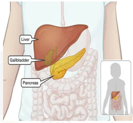
1. The Liver: The Body's Chemical Factory
The liver is the largest gland in the human body, situated in the upper right side of the abdominal cavity.
Bile Secretion: The liver produces Bile Juice, which is essential for the digestion of fats. It performs 'emulsification,' breaking down large fat globules into tiny droplets for enzymes to act upon.
Detoxification: It filters toxins from the blood and manages the storage of vitamins and minerals.
2. The Gallbladder: The Storage Unit
The gallbladder is a small, pear-shaped sac located just below the liver.
Function: It does not produce bile but serves as a storage and concentration chamber for it.
Release: When fatty food enters the small intestine, the gallbladder contracts and releases concentrated bile into the Duodenum via the common bile duct.
3. The Pancreas: The Dual-Function Gland
Located just below the stomach, the pancreas is a unique gland that acts as both an exocrine and endocrine organ.
Exocrine Function: It secretes Pancreatic Juice, which contains enzymes (like Trypsin and Lipase) to digest proteins, fats, and carbohydrates.
Endocrine Function: Specialized cells (Islets of Langerhans) secrete hormones directly into the blood:
Insulin: Lowers blood sugar levels.
Glucagon: Raises blood sugar levels by signaling the liver to release stored glucose.
| Organ | Secretion | Primary Target / Role |
|---|---|---|
| Liver | Bile Juice | Emulsification of Fats |
| Gallbladder | Stored Bile | Concentration and timely release of bile |
| Pancreas | Pancreatic Juice | Digestive enzymes for Carbs, Fats, Proteins |
| Pancreas | Insulin | Lowers Blood Glucose |
| Pancreas | Glucagon | Increases Blood Glucose |
Prelims tips
- Bile: Remember it contains no enzymes but is still vital for digestion.
- Largest Gland: Liver.
- Insulin/Glucagon: These are antagonistic hormones (work opposite to each other).
- Common Bile Duct: Formed by the joining of the cystic duct (from gallbladder) and hepatic duct (from liver).
Mains points
- Endocrine Regulation: The balance between Insulin and Glucagon is the cornerstone of metabolic homeostasis; disruption leads to lifestyle diseases like Type-2 Diabetes.
- Pathology: Gallstones occur when substances in bile (like cholesterol) crystallize, blocking the flow and causing severe pain and digestive issues.
- Public Health: Hepatitis (inflammation of the liver) and Fatty Liver disease are rising health concerns in India, linked to viral infections and dietary habits.
- Metabolic Processing: The liver's role in processing nutrients absorbed from the small intestine (via the portal vein) makes it the primary gatekeeper for systemic nutrition.
Did you know?
- The liver is the only organ in the human body that can completely regenerate itself even if only 25% of the original tissue remains.
- Bile juice is actually what gives feces its brown color; without it, waste would appear grey or clay-colored.
- The pancreas is about 6 inches long and shaped like a flat pear or a fish.
Respiratory System: The Mechanism of Gas Exchange
The Respiratory System is the biological network responsible for the intake of Oxygen (O2) and the removal of Carbon Dioxide (CO2). This continuous exchange is facilitated by the Alveoli in the lungs and the iron-rich protein Haemoglobin in the blood, ensuring that every cell in the body receives the energy needed for metabolism.
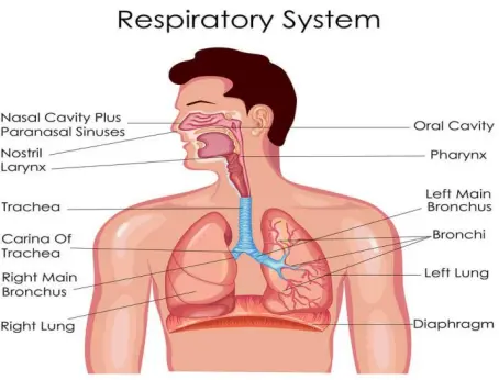
1. Breathing Mechanism
Breathing involves two main phases: Inspiration (inhalation) and Expiration (exhalation). It is driven by pressure gradients created by the diaphragm and intercostal muscles.
Inhalation: The diaphragm contracts and moves downward, increasing the volume of the chest cavity and pulling air into the lungs.
Exhalation: The diaphragm relaxes and moves upward, pushing CO2-rich air out of the lungs.
2. Alveoli: The Primary Exchange Site
The lungs contain millions of tiny, balloon-like structures called Alveoli. These are the actual sites where gas exchange occurs between the air and the blood.
Diffusion: O2 from the inhaled air diffuses across the thin alveolar membrane into the surrounding capillaries, while CO2 moves from the blood into the alveoli to be exhaled.
Surface Area: The vast number of alveoli provides a massive surface area to maximize the efficiency of this exchange.
3. Gas Transport: The Role of Haemoglobin
Once oxygen enters the blood, it must be transported to distant tissues.
Haemoglobin (Hb): A red-colored, iron-containing pigment in Red Blood Cells (RBCs). One Hb molecule can bind with up to four molecules of O2.
Oxyhaemoglobin: Oxygen binds with Hb to form oxyhaemoglobin, which travels through the arteries to reach body cells.
CO2 Transport: While some CO2 is carried by Hb, the majority is transported dissolved in the blood plasma as bicarbonate ions.
| Gas | Direction of Movement | Primary Carrier |
|---|---|---|
| Oxygen (O2) | Alveoli to Blood | Haemoglobin (RBCs) |
| Carbon Dioxide (CO2) | Blood to Alveoli | Plasma (as Bicarbonate) |
Prelims tips
- Alveoli: The structural and functional unit of the lungs.
- Haemoglobin: An iron-rich protein; deficiency leads to Anemia, which reduces oxygen-carrying capacity.
- Diaphragm: The primary muscle of respiration.
- Diffusion: The physical process (passive transport) by which gases move across the alveolar membrane.
Mains points
- Public Health: Air pollution and smoking cause chronic damage to alveoli (Emphysema), reducing the surface area available for gas exchange and leading to respiratory failure.
- High Altitude Adaptation: At high altitudes where O2 pressure is low, the body compensates by increasing RBC production and heart rate.
- Clinical Significance: Pulse oximetry measures the percentage of oxygen-saturated haemoglobin, a critical indicator during respiratory distress (like in COVID-19).
- Biological Efficiency: The thinness of the alveolar-capillary membrane is a prime example of biological engineering for rapid diffusion.
Did you know?
- Your left lung is slightly smaller than your right lung to make room for your heart!
- The total surface area of all the alveoli in an adult human's lungs is roughly the size of a tennis court.
- A person typically breathes about 12 to 16 times per minute while at rest.
Respiratory Disorders: Causes and Clinical Impact
Respiratory disorders encompass a range of conditions that impair the body's ability to exchange gases effectively. These range from chronic inflammatory conditions like Asthma to lifestyle-induced damage such as Emphysema. For UPSC, understanding the physiological changes in the lungs—specifically the bronchi and alveoli—and the impact of environmental pollutants is critical for Health and Environment modules.
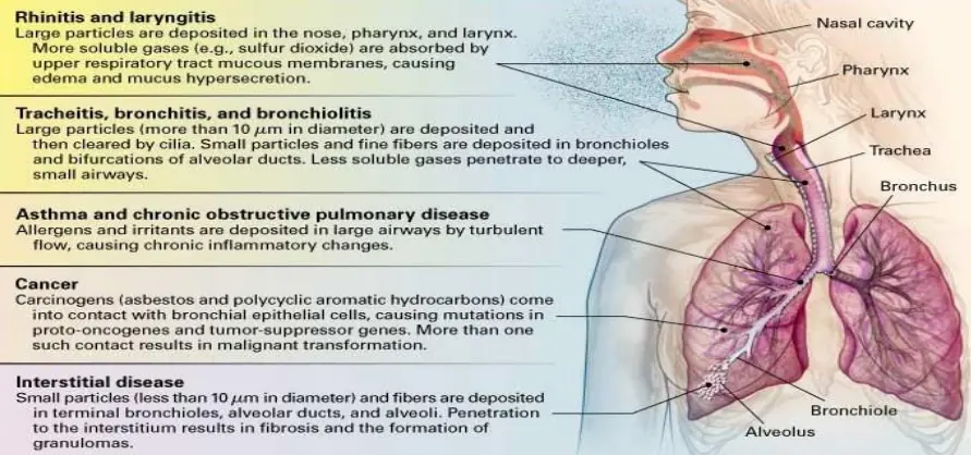
1. Chronic Respiratory Conditions
These disorders typically involve long-term inflammation or structural damage to the airway.
Asthma: A chronic disease characterized by the inflammation of the Bronchi and Bronchioles. It causes the airways to narrow and produce extra mucus, leading to difficulty in breathing, wheezing, and coughing.
Emphysema: A chronic disorder where the Alveolar walls are damaged. This reduces the surface area available for gas exchange. One of the major causes of this condition is cigarette smoking.
2. Occupational Respiratory Disorders
Many respiratory issues are caused by long-term exposure to industrial pollutants.
Mechanism: In certain industries involving stone-breaking or grinding, so much dust is produced that the body's defense mechanism cannot fully cope. Prolonged exposure leads to Fibrosis (proliferation of fibrous tissues).
Examples: Silicosis (caused by silica dust) and Asbestosis (caused by asbestos fibers), which result in serious lung damage.
3. Infections and Modern Challenges
Bronchitis: Inflammation of the lining of the bronchial tubes, which carry air to and from the lungs. It can be acute (often following a cold) or chronic.
COVID-19: A viral infection caused by SARS-CoV-2 that primarily attacks the respiratory system. It can lead to Pneumonia, where alveoli fill with fluid, and in severe cases, Acute Respiratory Distress Syndrome (ARDS) or multi-organ failure.
| Disorder | Affected Part | Primary Cause / Feature |
|---|---|---|
| Asthma | Bronchi & Bronchioles | Allergens, Inflammation, Wheezing |
| Emphysema | Alveolar Walls | Cigarette Smoking, Reduced surface area |
| Silicosis | Lung Tissue (Fibrosis) | Inhalation of Silica dust (Industrial) |
| Pneumonia | Alveoli | Infection leading to fluid accumulation |
Prelims tips
- Emphysema: Key keyword is 'damage to alveolar walls' and 'cigarette smoking'.
- Fibrosis: Proliferation of fibrous tissues, common in occupational diseases.
- Alveoli: The primary site of gas exchange; most disorders target this area to reduce O2 intake.
- Haemoglobin: Respiratory disorders often lead to lower oxygen saturation (SpO2) in the blood.
Mains points
- Environmental Policy: The rising incidence of Asthma and COPD in Indian cities underscores the need for stricter implementation of the National Clean Air Programme (NCAP).
- Occupational Safety: Strengthening labor laws and mandatory protective gear in mining and construction sectors are essential to prevent Silicosis and Asbestosis.
- Public Health: Smoking cessation programs and higher taxation on tobacco products are direct interventions to reduce the burden of Emphysema and Lung Cancer.
- Post-Pandemic Healthcare: The long-term impact of COVID-19 on lung health (Long COVID) requires specialized respiratory rehabilitation infrastructure.
Did you know?
- Cigarette smoke contains over 7,000 chemicals, many of which directly dissolve the delicate membranes of your alveoli, leading to permanent Emphysema.
- Your lungs are the only organs in your body that can float on water because they contain millions of air-filled alveoli!
- Laughter is actually a great exercise for the respiratory system as it forces the lungs to clear out 'stale' air and take in more oxygen.
Circulatory System: The Human Heart and Blood Flow
The Circulatory System, discovered by William Harvey, is the body's primary transport network. It delivers oxygen and nutrients to cells while removing waste products. In humans, the Heart acts as a muscular pump, ensuring Double Circulation—a specialized process that keeps oxygenated and deoxygenated blood separate for maximum efficiency.
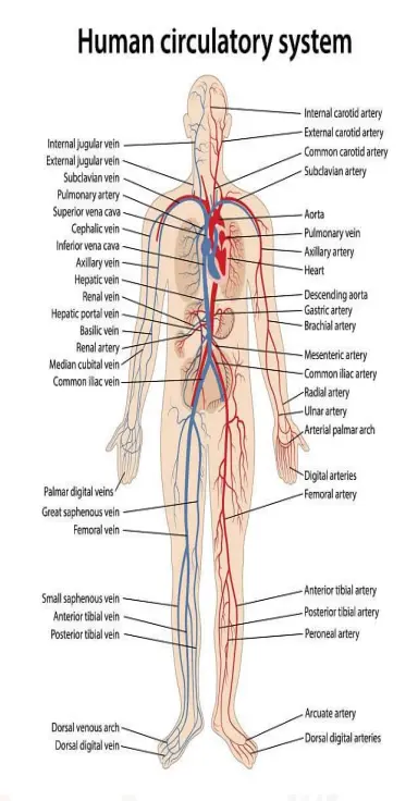
1. Structure of the Human Heart
The human heart is a four-chambered muscular organ roughly the size of a closed fist, protected by a double-walled membrane called the pericardium.
Atria (Auricles): Two upper, smaller chambers that receive blood.
Ventricles: Two lower, larger chambers with thick muscular walls that pump blood out to the lungs and body.
Valves: Structures like the Tricuspid and Bicuspid (Mitral) valves ensure that blood flows in only one direction, preventing backflow.
2. The Cardiac Cycle and Blood Pressure
The rhythmic contraction and relaxation of the heart constitute the cardiac cycle.
Heartbeat: A normal heart beats about 72 times per minute. Each cycle includes Systole (contraction) and Diastole (relaxation).
Cardiac Output: The heart pumps about 5 liters of blood per minute in a healthy adult.
Blood Pressure: It is the pressure exerted by blood against the walls of arteries. Normal BP is 120/80 mmHg.
3. Double Circulation and Blood Vessels
Humans possess double circulation, meaning blood passes through the heart twice for each complete circuit of the body.
Pulmonary Circulation: Deoxygenated blood travels from the heart to the lungs and returns oxygenated.
Systemic Circulation: Oxygenated blood travels from the heart to the rest of the body and returns deoxygenated.
Arteries vs. Veins: Arteries carry oxygen-rich blood away from the heart (high pressure), while veins carry oxygen-poor blood toward the heart (lower pressure, possess valves).
| Condition | Description | Impact |
|---|---|---|
| Hypertension | Blood pressure consistently above 140/90 | Damages brain and kidneys |
| Angina Pectoris | Acute chest pain due to low oxygen | Warning sign of heart issues |
| Heart Failure | Heart doesn't pump enough blood | Leads to fluid buildup in lungs |
| Atherosclerosis | Deposition of fats/cholesterol in arteries | Narrows the blood pathway |
Prelims tips
- William Harvey: Discovered the blood circulation system.
- SA Node: Known as the 'Natural Pacemaker', located in the right atrium.
- Arteries vs Veins: Pulmonary artery is the ONLY artery carrying deoxygenated blood; Pulmonary vein is the ONLY vein carrying oxygenated blood.
- Heart Sounds: 'Lub' is the closure of AV valves; 'Dub' is the closure of semilunar valves.
Mains points
- Non-Communicable Diseases (NCDs): Cardiovascular diseases (CVDs) are the leading cause of death globally, necessitating focus on preventive lifestyle changes.
- Public Health: The National Program for Prevention and Control of Cancer, Diabetes, Cardiovascular Diseases and Stroke (NPCDCS) in India targets early screening of hypertension.
- Technological Advancements: The use of Artificial Hearts and Stents represents the pinnacle of biomedical engineering in treating coronary artery diseases.
- Biological Integration: The close link between the respiratory and circulatory systems (Pulmonary circuit) highlights how the body optimizes gas transport for aerobic respiration.
Did you know?
- Your heart creates enough pressure to squirt blood up to 30 feet!
- The 'lub-dub' sound of a heartbeat is actually the sound of heart valves closing.
- If you stretched out all the blood vessels in an adult body, they would wrap around the Earth more than twice!
The Human Heart: Chambers, Rhythms, and Sounds
The human heart is a sophisticated muscular pump designed for maximum efficiency. With its Four-Chambered structure, it ensures a continuous flow of blood, moving roughly 5 liters every minute. Key clinical indicators like Heart Rate, Stroke Volume, and the distinctive Lub-Dub sounds provide a window into the health of the entire cardiovascular system.
1. Anatomy of the Heart Chambers
The human heart is divided into four distinct chambers to prevent the mixing of oxygenated and deoxygenated blood.
Atria (Upper Chambers): The right atrium receives deoxygenated blood from the body, while the left atrium receives oxygenated blood from the lungs. They have relatively thin walls.
Ventricles (Lower Chambers): The right ventricle pumps blood to the lungs, and the left ventricle pumps blood to the entire body. Ventricles have much thicker muscular walls, especially the left ventricle, to generate the pressure needed for systemic circulation.
2. Heart Rate and Cardiac Output
The rhythmic activity of the heart is regulated by internal electrical impulses.
Heart Rate: In a normal, resting adult, the heart beats approximately 70 to 75 times per minute (average 72 bpm).
Stroke Volume: During each cardiac cycle (one beat), each ventricle pumps out approximately 70 mL of blood. This is known as the stroke volume.
Cardiac Output: By multiplying the heart rate by the stroke volume (72 beats/min x 70 mL/beat), we find that the heart pumps about 5,000 mL (5 liters) of blood per minute.
3. The Music of the Heart: Lub and Dub
The heartbeat produces two characteristic sounds that can be heard through a stethoscope, caused by the closing of heart valves.
First Heart Sound (Lub): This is a low-pitched, longer sound associated with the closure of the Tricuspid and Bicuspid (Mitral) valves at the start of ventricular systole (contraction).
Second Heart Sound (Dub): This is a higher-pitched, shorter sound associated with the closure of the Semilunar valves at the beginning of ventricular diastole (relaxation).
| Parameter | Normal Value | Definition |
|---|---|---|
| Heart Rate | 72 beats/min | Number of heartbeats per minute |
| Stroke Volume | 70 mL | Volume of blood pumped per beat |
| Cardiac Output | ~5 Liters/min | Total volume of blood pumped per minute |
| Systole | Contraction phase | When heart pumps blood out |
| Diastole | Relaxation phase | When heart chambers fill with blood |
Prelims tips
- Double Circulation: Prevents mixing of oxygenated and deoxygenated blood; found in mammals and birds.
- Heart Sounds: Useful for clinical diagnosis; 'Lub' is S1, 'Dub' is S2.
- Formula: Cardiac Output = Heart Rate x Stroke Volume.
- Pacemaker: The SA Node initiates the cardiac cycle (located in the right atrium).
Mains points
- Technological Innovation: Understanding the cardiac cycle has led to the development of ECG (Electrocardiogram), which is essential for diagnosing arrhythmias and heart attacks.
- Public Health Burden: Cardiovascular diseases (CVDs) are the leading cause of death in India. Monitoring heart rate and BP is a primary preventive measure in primary healthcare.
- Exercise Physiology: Athletes have a lower resting heart rate (Bradycardia) but a much higher stroke volume, showcasing the heart's ability to adapt to physical stress.
- Biological Engineering: The unidirectional valves of the heart are a perfect example of how structure supports function by preventing backflow in a high-pressure system.
Did you know?
- A woman's heart typically beats slightly faster than a man's heart.
- The left ventricle is three times thicker than the right ventricle because it has to pump blood to your toes and head against gravity.
- Your heart does enough work in one day to drive a truck for 32 kilometers (20 miles).
Excretory System: Filtration and Waste Removal
The Excretory System is the body's purification plant, responsible for removing toxic metabolic wastes such as urea and ammonia. By maintaining Homeostasis—the balance of water and salts—it ensures the internal environment remains stable. The Kidneys are the primary organs here, acting as sophisticated biological filters that process hundreds of liters of blood daily.
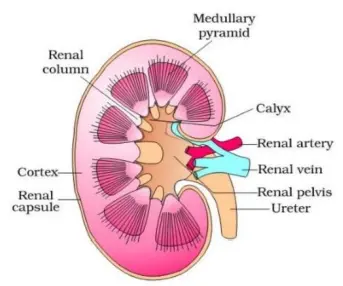
1. Role of the Excretory System
Metabolism produces various nitrogenous wastes that can be toxic if allowed to accumulate. The excretory system filters these from the blood and eliminates them from the body.
Metabolic Wastes: Includes Nitrogenous wastes (Urea, Uric Acid, Ammonia), excess salts, and water.
Primary Organs: A pair of bean-shaped Kidneys located on either side of the backbone.
2. Kidney Function: The Filtration Process
The kidneys filter the entire blood volume many times a day to maintain chemical balance.
Nephrons: Each kidney contains about a million functional units called Nephrons. These are the actual sites where filtration occurs.
Glomerulus: A cluster of capillaries where high-pressure filtration pushes water and waste out of the blood into the tubule.
Reabsorption: As the filtered fluid moves through the tubule, the body 'reclaims' useful substances like glucose, amino acids, and most of the water.
3. The Pathway of Urine
Once the waste is concentrated into urine, it follows a specific exit route:
Ureters: Two thin tubes that carry urine from the kidneys to the urinary bladder.
Urinary Bladder: A muscular sac that stores urine temporarily.
Urethra: The tube through which urine is finally expelled from the body.
| Organ | Function |
|---|---|
| Kidneys | Filter blood and produce urine |
| Ureters | Transport urine from kidney to bladder |
| Urinary Bladder | Temporary storage of urine |
| Urethra | Passage for urine expulsion |
Prelims tips
- Nephron: The structural and functional unit of the kidney.
- Urea: Produced in the Liver but excreted by the Kidney.
- Micturition: The clinical term for the act of passing urine.
- Homeostasis: Maintenance of a constant internal environment (water and salt balance).
Mains points
- Clinical Advancements: When kidneys fail, Dialysis (Artificial Kidney) is used to perform the filtration process, highlighting the vital role of osmotic balance.
- Public Health: High salt intake and dehydration are major causes of Kidney Stones and Chronic Kidney Disease (CKD) in India.
- Biological Regulation: The hormone ADH (Antidiuretic Hormone) controls how much water the kidneys reabsorb, directly linking the nervous system to excretion.
- Organ Donation: Kidney transplants are the most common form of organ transplant surgery, saving thousands from terminal renal failure.
Did you know?
- Your kidneys filter about 180 liters of blood every single day, but you only produce about 1.5 liters of urine!
- A person can live a perfectly healthy life with only one functioning kidney.
- If the nephrons from both kidneys were uncoiled and laid end-to-end, they would stretch for over 16 kilometers (10 miles).
Nephron: The Microscopic Filter and Urine Formation
The Nephron is the structural and functional unit of the kidney, acting as a complex miniature processing plant. Urine formation is not just a simple strain; it is a three-step high-precision process involving Filtration, Reabsorption, and Secretion. This ensures the body retains vital nutrients while precisely maintaining the Glomerular Filtration Rate (GFR) to stabilize internal fluid balance.
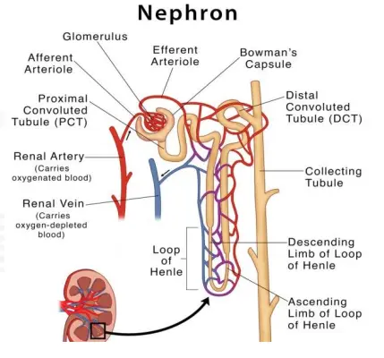
1. The Nephron Structure
Each kidney houses nearly one million nephrons. A nephron consists of two main parts: the Glomerulus and the Renal Tubule.
Glomerulus: A tuft of capillaries where the blood is first filtered under high pressure.
Bowman’s Capsule: A cup-like structure that encloses the glomerulus and collects the initial filtrate.
2. The Three Steps of Urine Formation
Urine is produced through a highly regulated sequence of events:
1. Glomerular Filtration: Blood is filtered through the glomerular membrane. About 1100-1200 ml of blood is filtered by the kidneys per minute.
2. Reabsorption: Nearly 99% of the filtrate is reabsorbed by the renal tubules. Essential substances like glucose, amino acids, and water are taken back into the blood.
3. Secretion: The tubular cells secrete substances like $H^+$ and $K^+$ into the filtrate to maintain the body's ionic and acid-base balance.
3. Glomerular Filtration Rate (GFR)
GFR is the amount of filtrate formed by the kidneys per minute.
Normal Value: In a healthy individual, the GFR is approximately 125 ml/minute, which totals about 180 liters of filtrate per day.
Regulation: The body has built-in mechanisms (like the Juxtaglomerular apparatus) to regulate GFR, ensuring the kidneys don't under-filter or over-filter regardless of blood pressure changes.
| Stage | Process Location | Key Activity |
|---|---|---|
| Filtration | Glomerulus | Filtering blood into the tubule |
| Reabsorption | Renal Tubules (PCT/Loop) | Reclaiming glucose, water, salts |
| Secretion | Renal Tubules (DCT) | Adding waste like $H^+$ ions to urine |
Prelims tips
- Nephron: Unit of Kidney. (Don't confuse with Neuron - Unit of Brain).
- GFR: Normal is 125 ml/min. A lower GFR indicates poor kidney health.
- 99%: The percentage of primary filtrate that is reabsorbed.
- Nitrogenous Waste: Urea is the main waste removed during this process.
Mains points
- Clinical Significance: Chronic Kidney Disease (CKD) is often staged based on GFR values, emphasizing its role as a primary indicator of renal efficiency.
- Homeostatic Balance: The 'Secretion' phase of urine formation is vital for blood pH regulation, highlighting the kidney's role as a metabolic buffer.
- Medical Innovation: Hemodialysis machines mimic the nephron's filtration and reabsorption processes to treat patients with end-stage renal disease.
- Physiological Adaptability: The nephron's ability to selectively reabsorb water is controlled by hormones like Vasopressin (ADH), showing the integration of the endocrine and excretory systems.
Did you know?
- Even though your kidneys create 180 liters of filtrate every day, 178.5 liters are reabsorbed, leaving only 1.5 liters to be excreted as urine.
- The Loop of Henle in the nephron is what allows mammals to produce concentrated urine, helping us conserve water.
- If your GFR drops significantly, it is a clinical sign of kidney failure or chronic kidney disease.
Excretory System: Filtration and Waste Removal
The Excretory System is the body's purification plant, responsible for removing toxic metabolic wastes such as urea and ammonia. By maintaining Homeostasis—the balance of water and salts—it ensures the internal environment remains stable. The Kidneys are the primary organs here, acting as sophisticated biological filters that process hundreds of liters of blood daily.
1. Role of the Excretory System
Metabolism produces various nitrogenous wastes that can be toxic if allowed to accumulate. The excretory system filters these from the blood and eliminates them from the body.
Metabolic Wastes: Includes Nitrogenous wastes (Urea, Uric Acid, Ammonia), excess salts, and water.
Primary Organs: A pair of bean-shaped Kidneys located on either side of the backbone.
2. Kidney Function: The Filtration Process
The kidneys filter the entire blood volume many times a day to maintain chemical balance. Each kidney contains about a million functional units called Nephrons.
Glomerulus: A cluster of capillaries where high-pressure filtration pushes water and waste out of the blood into the tubule.
Reabsorption: As the filtered fluid moves through the tubule, the body 'reclaims' useful substances like glucose, amino acids, and most of the water.
3. The Pathway of Urine
Once the waste is concentrated into urine, it follows a specific exit route:
Ureters: Two thin tubes that carry urine from the kidneys to the urinary bladder.
Urinary Bladder: A muscular sac that stores urine temporarily.
Urethra: The tube through which urine is finally expelled from the body.
| Organ | Function |
|---|---|
| Kidneys | Filter blood and produce urine |
| Ureters | Transport urine from kidney to bladder |
| Urinary Bladder | Temporary storage of urine |
| Urethra | Passage for urine expulsion |
Prelims tips
- Nephron: The structural and functional unit of the kidney.
- Urea: Produced in the Liver but excreted by the Kidney.
- Micturition: The clinical term for the act of passing urine.
- Homeostasis: Maintenance of a constant internal environment (water and salt balance).
Mains points
- Clinical Advancements: When kidneys fail, Dialysis (Artificial Kidney) is used to perform the filtration process, highlighting the vital role of osmotic balance.
- Public Health: High salt intake and dehydration are major causes of Kidney Stones and Chronic Kidney Disease (CKD) in India.
- Biological Regulation: The hormone ADH (Antidiuretic Hormone) controls how much water the kidneys reabsorb, directly linking the nervous system to excretion.
- Organ Donation: Kidney transplants are the most common form of organ transplant surgery, saving thousands from terminal renal failure.
Did you know?
- Your kidneys filter about 180 liters of blood every single day, but you only produce about 1.5 liters of urine!
- A person can live a perfectly healthy life with only one functioning kidney.
- If the nephrons from both kidneys were uncoiled and laid end-to-end, they would stretch for over 16 kilometers (10 miles).
Metabolic Wastes: The Body's By-products
The human body produces various Metabolic Wastes as by-products of chemical reactions like digestion and cellular respiration. These include nitrogenous compounds like Urea, gases like CO2, and various ions. If not removed through specialized excretory pathways, these substances can become toxic, disrupting the body's internal chemical balance.
1. Primary Nitrogenous Wastes
Nitrogen-containing waste is produced during the breakdown of proteins and nucleic acids. They vary in toxicity and water requirement for removal:
Ammonia: The most toxic form; requires massive amounts of water for elimination (rare in humans, common in aquatic animals).
Urea: The main nitrogenous waste in humans. It is produced in the Liver and filtered out by the Kidneys.
Uric Acid: The least toxic form; requires very little water for removal.
2. Gaseous and Liquid Wastes
Apart from nitrogen, the body must also expel carbon and excess fluids.
Carbon Dioxide (CO2): A byproduct of cellular respiration, eliminated primarily by the Lungs.
Water: While essential, excess water is considered waste and is removed through urine, sweat, and breath.
3. Secondary Excretory Pathways
While kidneys are the 'star' filters, other organs assist in waste management:
Liver: Secretes Bile containing cholesterol, degraded steroid hormones, vitamins, and drugs for elimination via the digestive tract.
Skin: Sweat Glands remove water, NaCl, and small amounts of urea. Sebaceous Glands eliminate certain lipids.
Lungs: Remove nearly 18 liters of CO2 per hour and significant water vapor.
4. Ionic and Other Wastes
The body also regulates the levels of various ions which can become 'waste' if present in excess:
Ions: Sodium (Na+), Potassium (K+), Chloride (Cl-), Phosphates, and Sulphates.
Bile Pigments: Bilirubin and Biliverdin (from broken down RBCs), which give color to waste.
| Waste Product | Excretory Organ | Form of Removal |
|---|---|---|
| Urea | Kidneys | Urine |
| CO2 & Water Vapor | Lungs | Exhaled Air |
| Bile Pigments / Drugs | Liver | Feces (Digestive tract) |
| NaCl & Lactic Acid | Skin | Sweat |
| Sebum / Lipids | Skin | Secretions via Sebaceous Glands |
Prelims tips
- Urea Cycle: Occurs in the Liver (Ornithine cycle).
- Toxicity Rank: Ammonia > Urea > Uric Acid.
- Lungs: Not just for breathing; they remove 18L of CO2 per hour.
- Sweat: Contains NaCl, Lactic Acid, and small amounts of Urea.
Mains points
- Biological Adaptation: The shift from Ammonia to Urea/Uric acid excretion allowed vertebrates to move from water to land by conserving precious body fluids.
- Environmental Health: Heavy metal accumulation (like Lead or Mercury) in the liver and kidneys highlights the limits of the body's natural detoxification systems.
- Diagnostic Value: The presence of unusual wastes (like glucose or high protein) in urine is a primary clinical indicator of systemic diseases like Diabetes or Nephritis.
- Clinical Significance: Understanding the liver's role in drug metabolism is crucial for pharmacology and determining correct medicine dosages.
Did you know?
- The Liver doesn't just filter; it's a recycling center. It breaks down old Red Blood Cells to create bile pigments like Bilirubin.
- You lose about half a liter of water every day just by breathing!
- Sweat is 99% water; the 'stink' of sweat is actually caused by bacteria on the skin breaking down the 1% of urea and salts.
The Human Kidney: Structure and Vital Functions
The Kidneys are a pair of reddish-brown, bean-shaped organs located in the abdominal cavity. They serve as the body's primary filtration system, processing about 180 liters of blood daily to maintain Homeostasis. Beyond waste removal, they regulate blood pressure, electrolyte balance, and red blood cell production.
1. External and Internal Anatomy
Each kidney is about 10-12 cm long and is protected by a tough fibrous capsule.
Renal Cortex: The outer layer where the filtration units (nephrons) start.
Renal Medulla: The inner region divided into conical masses called medullary pyramids.
Renal Pelvis: A funnel-shaped space that collects urine before it travels to the ureters.
2. The Nephron: The Functional Unit
Each kidney contains nearly one million Nephrons. A nephron is a microscopic structure where the actual work of blood purification happens.
Glomerulus: A cluster of capillaries that filters blood under high pressure.
Bowman’s Capsule: The cup-like start of the nephron that catches the filtrate.
Renal Tubules: Long tubes where reabsorption of water, glucose, and essential salts occurs.
3. Key Functions of the Kidney
The kidneys are far more than just 'waste filters'; they are chemical regulators:
Excretion: Removal of nitrogenous wastes like urea, uric acid, and ammonia.
Osmoregulation: Maintaining the balance of water and salts (electrolytes) in the blood.
Blood Pressure Regulation: Through the release of the enzyme Renin.
Hormone Production: Secretion of Erythropoietin (stimulates RBC production) and activation of Vitamin D.
| Parameter | Normal Value | Significance |
|---|---|---|
| GFR | 125 ml/min | Measures filtration efficiency |
| Urea Output | ~25-30 g/day | Primary nitrogenous waste removed |
| Urine pH | 6.0 (Average) | Maintains acid-base balance |
| Daily Urine Volume | 1 - 1.5 Liters | Indicator of hydration and kidney health |
Prelims tips
- Erythropoietin: The kidney hormone responsible for stimulating Red Blood Cell production in bone marrow.
- Renal Calculus: The scientific name for Kidney Stones, often caused by calcium oxalate crystals.
- Hemodialysis: An artificial process to filter blood when kidneys fail.
- Micturition: The reflex process of passing urine.
Mains points
- Lifestyle Impact: The rise of Chronic Kidney Disease (CKD) in India is heavily linked to secondary epidemics of Diabetes and Hypertension.
- Ethical Medicine: Organ transplantation policies (Transplantation of Human Organs Act) are critical to curb the illegal trade of kidneys.
- Biotechnology: Advances in 'Bio-artificial Kidneys' and 3D organ printing are emerging solutions to the shortage of kidney donors.
- Environmental Toxins: Exposure to heavy metals (Lead, Cadmium) and certain pesticides can lead to nephrotoxicity, highlighting a link between environmental health and kidney failure.
Did you know?
- Even though kidneys represent only 0.5% of your body weight, they receive 20-25% of the total blood pumped by the heart every minute.
- The right kidney is usually slightly lower than the left to accommodate the large space occupied by the liver.
- A healthy human can live a long and normal life with only one kidney; it will simply grow larger to do the work of two.
Urine Formation: The Three-Step Renal Process
Urine formation is a highly precise physiological process conducted by the Nephrons to maintain the body's homeostatic balance. It involves filtering the entire blood volume to remove nitrogenous wastes while strategically reclaiming essential water and nutrients. This process is governed by three distinct stages: Glomerular Filtration, Reabsorption, and Secretion.
1. Glomerular Filtration (Ultrafiltration)
This is the first step where blood is filtered under high pressure in the Glomerulus.
Mechanism: Blood enters the glomerulus through the afferent arteriole. Because the outgoing vessel is narrower, high pressure is created, forcing water and small solutes out of the blood and into the Bowman’s Capsule.
The Filtrate: The resulting fluid (glomerular filtrate) contains water, glucose, amino acids, and urea, but is free of blood cells and large proteins.
GFR: The volume of filtrate formed by both kidneys per minute is the Glomerular Filtration Rate, which is normally about 125 ml/min.
2. Tubular Reabsorption
As the filtrate travels through the renal tubule, the body 'reclaims' substances it cannot afford to lose.
Volume Reduction: Nearly 99% of the 180 liters of daily filtrate is reabsorbed back into the blood.
Selective Uptake: In the Proximal Convoluted Tubule (PCT), almost all glucose and amino acids are reabsorbed. Water and electrolytes (Na+, Cl-) are reabsorbed throughout the tubule, especially in the Loop of Henle and Collecting Duct.
3. Tubular Secretion
This is the final 'fine-tuning' stage where the body actively removes specific ions and wastes from the blood into the tubule.
Function: It helps in maintaining the Ionic balance and pH of the body fluids.
Substances: Hydrogen ions (H+), Potassium ions (K+), and ammonia are secreted into the filtrate.
Result: The final concentrated fluid, now called Urine, moves into the collecting duct and eventually to the urinary bladder.
| Step | Location | Primary Action |
|---|---|---|
| Filtration | Glomerulus | Non-selective pressure filtration of blood |
| Reabsorption | Renal Tubules | Selective recovery of water, glucose, and salts |
| Secretion | Distal Tubule | Active removal of H+, K+, and ammonia into urine |
Prelims tips
- GFR: Normal value is 125 ml/min or 180 liters/day.
- PCT: Site of maximum reabsorption (70-80% of electrolytes and water).
- ADH (Vasopressin): Hormone that increases water reabsorption in the collecting duct during dehydration.
- Micturition: The process of releasing urine from the bladder.
Mains points
- Clinical Correlation: Measuring GFR is the gold standard for assessing kidney health; a decline in GFR is the primary marker for Chronic Kidney Disease (CKD).
- Acid-Base Homeostasis: Through tubular secretion of H+ ions and reabsorption of bicarbonate, the kidneys act as a powerful buffer system for blood pH.
- Pharmacology: Many drugs (like antibiotics) are cleared from the body through 'Tubular Secretion', which explains why their dosage depends on renal efficiency.
- Evolutionary Adaptation: The ability of the human nephron to produce concentrated urine (via the Countercurrent Mechanism) allowed mammals to successfully inhabit arid terrestrial environments.
Did you know?
- Your kidneys filter enough blood every day to fill about 40 large gas tanks, but the amount of actual urine produced is only about 1.5 liters.
- Glucosuria (glucose in urine) is a classic indicator that the reabsorption capacity of the nephrons has been exceeded, often due to Diabetes.
- The Yellow color of urine comes from a pigment called Urochrome, which is produced from the breakdown of old hemoglobin.
Secondary Excretory Organs: Beyond the Kidneys
While the kidneys are the primary filtration units, they are assisted by other vital organs—the Lungs, Liver, and Skin. These organs help eliminate metabolic by-products like Carbon Dioxide, bile pigments, and excess salts, ensuring that the body's internal environment remains detoxified and balanced.
1. The Lungs: Gaseous Excretion
The lungs play a massive role in removing gaseous waste produced during cellular respiration.
Carbon Dioxide (CO2): Our lungs eliminate large amounts of CO2 (approximately 18 liters per hour).
Water Vapor: Significant amounts of water are also lost through the breath daily, especially in dry or cold environments.
2. The Liver: The Metabolic Filter
The liver is the largest gland and serves as a major site for breaking down complex toxins and old blood cells.
Bile Secretion: The liver secretes bile which contains substances like Bilirubin, Biliverdin (degraded hemoglobin), cholesterol, degraded steroid hormones, vitamins, and drugs.
Elimination Route: These substances ultimately pass out of the body along with digestive waste (feces).
3. The Skin: Sweat and Sebum
The skin eliminates waste through two types of glands, primarily aiding in temperature regulation and waterproofing.
Sweat Glands: Produce sweat, a watery fluid containing NaCl (salt), small amounts of Urea, and lactic acid. Its primary aim is cooling, but it serves an excretory function.
Sebaceous Glands: Eliminate certain substances like sterols, hydrocarbons, and waxes through sebum, providing a protective oily covering for the skin.
| Organ | Excretory Product | Form |
|---|---|---|
| Lungs | CO2 and Water vapor | Gas/Breath |
| Liver | Bile pigments, Cholesterol, Drugs | Bile/Feces |
| Skin (Sweat) | NaCl, Urea, Lactic Acid | Liquid (Sweat) |
| Skin (Sebaceous) | Sterols, Hydrocarbons, Waxes | Oily (Sebum) |
Prelims tips
- Liver: Site of urea synthesis (Ornithine cycle), though kidneys excrete it.
- Bilirubin/Biliverdin: These are waste products of hemoglobin breakdown excreted via bile.
- Sebum: Provides a protective, oily layer but also serves as an excretory medium for lipids.
- Lungs: Crucial for acid-base balance by regulating CO2 levels.
Mains points
- Detoxification Mechanisms: The liver's role in drug metabolism is critical for medical science; liver failure leads to the toxic accumulation of drugs and metabolic by-products.
- Homeostatic Integration: The coordination between lungs (CO2) and kidneys (Bicarbonate) is the body's primary defense against blood pH fluctuations.
- Clinical Significance: Skin conditions or excessive sweating (Hyperhidrosis) can sometimes indicate underlying metabolic or endocrine imbalances.
- Environmental Health: Exposure to fat-soluble toxins often impacts the liver's ability to secrete bile effectively, leading to secondary digestive and excretory issues.
Did you know?
- The Liver is often called the 'Master Chemist' because it prepares toxic substances like ammonia to be safely converted into urea for the kidneys to handle.
- Your skin is your largest organ, and while it isn't the 'main' filter, it can release about 1% of the body's urea through sweat.
- Bile pigments (Bilirubin) are what give human waste its characteristic brown color. Without the liver's excretory role, waste would look grey.
Kidney Disorders: Renal Impairment and Pathologies
Kidney disorders occur when the renal system fails to filter blood, maintain fluid balance, or regulate electrolytes effectively. These conditions range from the formation of hard mineral deposits known as Renal Calculi to the life-threatening state of Uremia, where urea accumulates in the blood. For UPSC, understanding the physiological basis of these disorders and the medical interventions like Hemodialysis is essential.
1. Uremia and Kidney Failure
Uremia is a serious condition resulting from the accumulation of urea in the blood due to malfunctioning kidneys.
Uremia: When the GFR drops significantly, nitrogenous wastes build up in the blood, which can lead to kidney failure.
Hemodialysis: In patients with kidney failure, blood is filtered artificially. Blood is drained from a convenient artery, mixed with an anticoagulant like Heparin, and passed through a cellophane tube in a dialyzing unit. This tube is porous and allows nitrogenous wastes to diffuse out into a dialyzing fluid while keeping plasma proteins and blood cells inside.
Kidney Transplantation: The ultimate method for the correction of acute renal failure is the transplantation of a functional kidney from a donor, preferably a close relative to minimize immune rejection.
2. Renal Calculi (Kidney Stones)
Renal calculi are hard deposits made of minerals and salts that form inside the kidneys.
Formation: Stones or insoluble masses of crystallized salts (usually Oxalates of calcium) form within the kidney.
Impact: They can cause severe pain (renal colic) and may obstruct the flow of urine in the ureter or renal pelvis.
3. Glomerulonephritis
This is a specific inflammatory condition affecting the filtration units of the kidney.
Definition: Inflammation of the Glomeruli of the kidney.
Causes: Often caused by an immune response to infections or autoimmune diseases, leading to blood or protein leaking into the urine (Hematuria/Proteinuria).
| Disorder | Site of Issue | Key Feature |
|---|---|---|
| Uremia | Bloodstream | Accumulation of Urea |
| Renal Calculi | Renal Pelvis/Ureter | Stone formation (Oxalates) |
| Glomerulonephritis | Glomerulus | Inflammation/Immune response |
| Glycosuria | Renal Tubules | Presence of Glucose in urine |
| Proteinuria | Filtration Membrane | Protein leaking into urine |
Prelims tips
- Heparin: Anticoagulant added to blood during dialysis to prevent clotting outside the body.
- Calcium Oxalate: The most common chemical compound found in kidney stones.
- Hematuria: Presence of blood in urine, often a sign of inflammation or stones.
- Uremia: Indicates that the kidneys are filtering less than 15% of the normal rate.
Mains points
- Ethical Concerns: The global shortage of organs leads to illegal 'kidney rackets,' highlighting the need for strict enforcement of the Transplantation of Human Organs Act (THOA).
- Lifestyle Disease Burden: Diabetes and Hypertension are the primary 'silent killers' leading to End-Stage Renal Disease (ESRD) in India.
- Technological Innovation: Wearable artificial kidneys and stem cell research for organ regeneration are the next frontiers in treating renal disorders.
- Public Health: Awareness about hydration and diet (limiting salt and oxalates) is the most cost-effective way to prevent the rising incidence of renal calculi.
Did you know?
- Kidney stones can be as small as a grain of sand or as large as a golf ball!
- The dialyzing fluid used in dialysis has the exact same composition as plasma, except it lacks nitrogenous wastes, allowing waste to move out of the blood via simple diffusion.
- A person on dialysis can filter their entire blood volume several times in a single 4-hour session.
Endocrine and Vertebral Systems: Regulation and Structure
The Endocrine System acts as the body's chemical communication network, using hormones to regulate metabolism, growth, and reproduction. In contrast, the Vertebral System (part of the Skeletal System) provides the physical framework and protection for the central nervous system. Together, they ensure the body is both structurally sound and chemically balanced.
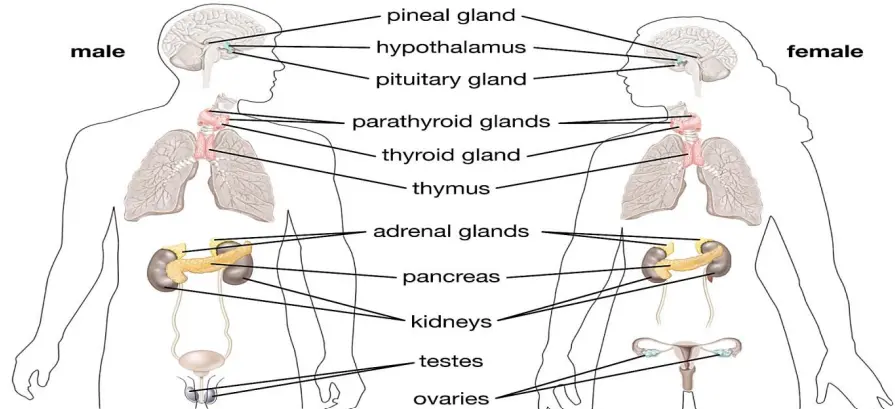
1. The Endocrine System (Chemical Messengers)
Unlike exocrine glands (like salivary glands), endocrine glands are ductless and release Hormones directly into the bloodstream.
Hypothalamus: The master controller; it secretes releasing hormones (like GnRH, TRH) that regulate the pituitary gland.
Pituitary Gland: Known as the 'Master Gland', it produces Growth Hormone (GH), TSH, and LH/FSH.
Thyroid Gland: Produces Thyroxine, which regulates the basal metabolic rate (BMR). It requires iodine for synthesis.
Adrenal Glands: Located atop the kidneys, they release Adrenaline (Epinephrine) for 'Fight or Flight' responses.
Pancreas: A composite gland. Insulin lowers blood sugar, while Glucagon raises it.
2. The Vertebral System (Structural Support)
The vertebral column (backbone) is a key part of the human axial skeleton, consisting of 26 serially arranged units called vertebrae.
Function: It protects the spinal cord, supports the head, and serves as the point of attachment for ribs and musculature.
Regions:
Cervical (7): Neck region.
Thoracic (12): Mid-back, attached to ribs.
Lumbar (5): Lower back, bears the most weight.
Sacrum & Coccyx: Fused vertebrae at the base.
3. Hormones by Chemical Nature
Hormones are classified based on their chemical structure:
Proteins/Peptides: Insulin, Glucagon, Pituitary hormones.
Steroids: Cortisol, Testosterone, Estradiol, Progesterone.
Iodothyronines: Thyroid hormones.
Amino-acid derivatives: Epinephrine (Adrenaline).
| Gland | Key Hormone | Primary Function |
|---|---|---|
| Pituitary | Growth Hormone | Body growth and development |
| Thyroid | Thyroxine | Regulates Metabolism |
| Adrenal | Adrenaline | Emergency response (Fight/Flight) |
| Pancreas | Insulin | Lowers Blood Glucose |
| Thymus | Thymosins | Immune system development |
Prelims tips
- Exocrine vs Endocrine: Exocrine has ducts (Saliva, Sweat); Endocrine is ductless (Hormones).
- Iodine: Essential for Thyroid health; deficiency leads to Goitre.
- Insulin: Produced by Beta cells of Pancreas; deficiency causes Diabetes Mellitus.
- Vertebral Formula: $C7, T12, L5, S1, Co1$ (Total 26 in adults).
Mains points
- Lifestyle Diseases: Hormonal imbalances due to stress and diet are leading to a surge in PCOD, Thyroid issues, and Diabetes in India.
- Ergonomics and Health: Sedentary lifestyles are causing an increase in vertebral disorders like Spondylitis and Slip Disc, impacting workforce productivity.
- Regulatory Feedback: The endocrine system works on a 'negative feedback mechanism'—when hormone levels are high, the body signals to stop production to maintain balance.
- Evolutionary Biology: The transition to a 'Bipedal' (two-legged) walk in humans led to the 'S' shaped curve of the vertebral column to balance the body's weight.
Did you know?
- The Pituitary gland is only about the size of a pea, yet it controls most other endocrine glands in the body.
- Humans and giraffes both have exactly 7 cervical (neck) vertebrae, even though a giraffe's neck is much longer!
- Your Adrenal glands can trigger a strength boost so high during emergencies that people have been known to lift cars to save others.
The Human Brain: Command Center of the Body
The Human Brain is the most complex organ, acting as the central command center for the nervous system. It processes sensory information, regulates blood pressure and breathing, and enables cognitive functions like thinking, memory, and emotion. Structurally, it is divided into three primary regions: the Forebrain, Midbrain, and Hindbrain, each housing specialized centers for vital life processes.
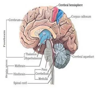
1. Anatomy of the Brain
The brain is protected by the skull (cranium) and cushioned by cerebrospinal fluid. It is divided into three major parts:
Forebrain (Prosencephalon): The largest part. It includes the Cerebrum (seat of intelligence), Thalamus (sensory relay), and Hypothalamus (controls body temperature, hunger, and thirst).
Midbrain (Mesencephalon): Located between the forebrain and hindbrain. it acts as a relay station for auditory and visual signals.
Hindbrain (Rhombencephalon): Consists of the Pons, Cerebellum (maintains balance and posture), and Medulla Oblongata (controls involuntary actions like heartbeat and respiration).
2. The Cerebrum and Hemispheres
The Cerebrum is divided into two halves called left and right cerebral hemispheres, connected by a tract of nerve fibers called the Corpus Callosum.
Frontal Lobe: Reasoning, planning, and speech.
Parietal Lobe: Movement, orientation, and recognition.
Occipital Lobe: Visual processing.
Temporal Lobe: Auditory perception and memory.
3. The Brain Stem
The midbrain and hindbrain (excluding the cerebellum) form the brain stem.
Function: It connects the brain to the spinal cord.
Life Support: It houses centers that regulate critical involuntary functions such as breathing, swallowing, and heart rate.
| Region | Specific Part | Main Function |
|---|---|---|
| Forebrain | Cerebrum | Intelligence, Memory, Logic |
| Forebrain | Hypothalamus | Thermoregulation, Hunger, Thirst |
| Hindbrain | Cerebellum | Precision, Balance, Posture |
| Hindbrain | Medulla | Respiration, Blood Pressure, Gastric secretion |
Prelims tips
- Hypothalamus: Known as the 'thermostat' of the body; also regulates sleep cycles.
- Medulla Oblongata: Essential for life; injury to this part often results in instant death due to respiratory failure.
- Cerebellum: Alcohol affects this part first, which is why intoxicated people lose their balance.
- Grey Matter vs White Matter: Grey matter consists of nerve cell bodies; White matter consists of myelinated axons.
Mains points
- Neuroplasticity: The brain's ability to reorganize itself by forming new neural connections throughout life is vital for recovery from stroke and learning new skills.
- Mental Health: Chemical imbalances in neurotransmitters like Dopamine and Serotonin are linked to clinical depression and anxiety, highlighting the biological basis of mental health.
- Brain-Computer Interface (BCI): Modern technology is developing ways to link human thoughts directly to computers, potentially helping paralyzed individuals regain movement.
- Ethics of AI: As Artificial Intelligence mimics neural networks, understanding the human brain's architecture is essential for setting ethical and functional boundaries for machines.
Did you know?
- The human brain is 75% water and is the fattiest organ in the body, consisting of nearly 60% fat.
- Information in your brain travels at speeds of up to 430 kilometers per hour (268 mph).
- The brain itself cannot feel pain; even though it processes pain signals for the entire body, it lacks pain receptors.
The Neuron: Structural Unit of the Nervous System
A Neuron is a highly specialized microscopic cell responsible for transmitting electrical and chemical signals throughout the body. Known as the functional unit of the nervous system, it allows the brain to communicate with muscles and organs. Unlike most other cells, mature neurons generally do not divide, making their protection and health vital for lifelong cognitive function.
1. Structure of a Neuron
A typical neuron consists of three major parts: the Cell Body, Dendrites, and Axon.
Cell Body (Soma): Contains the nucleus and typical cell organelles. It also has specific granular bodies called Nissl's granules.
Dendrites: Short, branching fibers that project out of the cell body. They receive incoming signals and transmit them toward the cell body.
Axon: A long fiber that transmits impulses away from the cell body to a synapse or neuromuscular junction.
Myelin Sheath: An insulating layer (formed by Schwann cells) that wraps around the axon to increase the speed of signal transmission.
2. Types of Neurons
Based on their function and the number of processes, neurons are categorized as:
Sensory Neurons: Carry impulses from sensory receptors (like skin or eyes) toward the Central Nervous System (CNS).
Motor Neurons: Carry impulses from the CNS to 'effector' organs like muscles and glands.
Interneurons: Located within the brain and spinal cord, they connect sensory and motor neurons.
3. Nerve Impulse Transmission
Signals travel through a neuron as electrical impulses but cross the gap between neurons chemically.
Synapse: The junction between the axon terminal of one neuron and the dendrite of the next.
Neurotransmitters: Chemical messengers (like Acetylcholine or Dopamine) released into the synaptic cleft to pass the signal to the next cell.
| Component | Function |
|---|---|
| Dendrites | Receive signals from other neurons |
| Axon | Conducts impulses away from the cell body |
| Myelin Sheath | Provides electrical insulation and speeds up impulses |
| Synaptic Knob | Releases neurotransmitters into the synapse |
| Nissl's Granules | Site of protein synthesis in the cell body |
Prelims tips
- Nissl's Granules: Unique to neurons, found in the cell body and dendrites but absent in the axon.
- Saltatory Conduction: The rapid jumping of electrical signals between 'Nodes of Ranvier' on myelinated axons.
- Synapse: Signals move in only one direction across a synapse.
- Neurotransmitters: Chemicals stored in synaptic vesicles at the axon terminal.
Mains points
- Degenerative Diseases: Conditions like Alzheimer's and Parkinson's involve the progressive loss of specific neurons, impacting memory and movement.
- Neural Plasticity: The brain's ability to strengthen or weaken synapses based on activity is the biological basis for learning and memory.
- Impact of Toxins: Many neurotoxins (like those in snake venom or certain pesticides) work by blocking the chemical receptors at the synapse, leading to paralysis.
- Artificial Intelligence: Artificial Neural Networks (ANNs) in computing are inspired by the structure and interconnectedness of biological neurons.
Did you know?
- Neurons are the longest cells in the human body; a single motor neuron can stretch from your lower spine all the way to your big toe!
- Unlike skin or bone cells, most neurons in the brain do not regrow if they die, which is why brain and spinal cord injuries are so serious.
- Your brain contains approximately 86 billion neurons, forming trillions of connections.
The Vertebral System: Structural Framework of the Body
The Vertebral Column, also known as the backbone or spine, is a primary component of the human axial skeleton. It consists of 26 serially arranged units called Vertebrae. This system is not just a rigid rod; it is a flexible, curved structure that supports the head, protects the spinal cord, and serves as the main attachment point for the ribs and back muscles.
1. Structure of a Vertebra
Each vertebra has a central hollow portion called the Neural Canal through which the spinal cord passes.
Intervertebral Discs: Between adjacent vertebrae, there are fibrocartilaginous pads that act as shock absorbers and allow for limited movement of the spine.
Articulation: The first vertebra is the Atlas, which articulates with the occipital condyles of the skull, allowing the 'nodding' motion of the head.
2. Regions of the Vertebral Column
The spine is divided into five distinct regions based on the location and structure of the vertebrae.
Cervical (7): Located in the neck region. All mammals, including giraffes and humans, almost always have seven cervical vertebrae.
Thoracic (12): Located in the upper/mid-back; these articulate with the ribs.
Lumbar (5): Located in the lower back; these are the largest and strongest as they bear the most weight.
Sacrum (1): A single triangular bone formed by the fusion of five sacral vertebrae in adults.
Coccyx (1): The 'tailbone,' formed by the fusion of four small vertebrae.
3. Natural Curvatures
A healthy adult spine has an 'S' shape when viewed from the side.
Function: These curves (Cervical, Thoracic, Lumbar, and Pelvic) help in balancing the body weight while standing upright, absorbing shocks during walking, and protecting the column from fractures.
| Region | Number of Vertebrae | Location |
|---|---|---|
| Cervical | 7 | Neck |
| Thoracic | 12 | Chest/Upper Back |
| Lumbar | 5 | Lower Back |
| Sacrum | 1 (5 fused) | Pelvic area |
| Coccyx | 1 (4 fused) | Tailbone |
Prelims tips
- Vertebral Formula: $C_7 T_{12} L_5 S_{(5)} Co_{(4)}$ for a total of 26 units in adults.
- Cervical Vertebrae: The presence of 7 cervical vertebrae is a characteristic feature of nearly all mammals.
- Neural Canal: The spinal cord passes through this central hollow portion.
- Atlas & Axis: The first and second vertebrae that allow for complex head movements.
Mains points
- Evolutionary Adaptation: The 'S' shaped curvature of the human spine is a direct result of 'Bipedalism' (walking on two legs), which requires a unique center of gravity.
- Public Health: Increasing cases of Spondylitis and Slip Disc in urban populations are attributed to sedentary lifestyles and poor ergonomics.
- Biological Engineering: The intervertebral discs are a marvel of nature, acting as hydraulic shock absorbers that protect the brain from impact during running and jumping.
- Surgical Advancements: Minimally invasive spinal surgeries (like Microdiscectomy) have transformed the treatment of chronic back pain and nerve compression.
Did you know?
- Humans are born with 33 individual vertebrae, but by the time we become adults, some fuse together to leave us with just 26.
- The Lumbar vertebrae are the largest because they have to support the entire weight of your upper body.
- You are actually slightly taller in the morning than at night because gravity compresses the cartilage discs in your spine throughout the day.
The Endocrine System: The Body's Chemical Messaging Network
The Endocrine System consists of ductless glands that secrete Hormones directly into the bloodstream to regulate distant target organs. Unlike the nervous system, which uses electrical impulses for rapid responses, the endocrine system uses chemical signals to manage long-term processes like growth, metabolism, and reproduction.
1. Major Endocrine Glands and Their Locations
Endocrine glands are scattered throughout the body, coordinated largely by the brain.
Hypothalamus: Located in the forebrain, it is the 'master of the master gland.' It links the nervous system to the endocrine system.
Pituitary Gland: A pea-sized gland at the base of the brain. It secretes hormones that control other glands (like TSH, LH, and FSH) and Growth Hormone (GH).
Thyroid Gland: Located in the neck, it produces Thyroxine, which regulates the Basal Metabolic Rate (BMR).
Adrenal Glands: Sitting atop the kidneys, they produce 'fight or flight' hormones like Adrenaline.
Pancreas: A dual-purpose (heterocrine) gland that regulates blood sugar via Insulin and Glucagon.
2. Mechanism of Hormone Action
Hormones act as primary messengers. They travel through the blood and only affect cells that have specific receptors for them.
Water-soluble hormones: (e.g., Insulin) Bind to receptors on the cell surface and trigger secondary messengers inside the cell.
Lipid-soluble hormones: (e.g., Steroids like Estrogen) Can pass through the cell membrane and interact directly with the cell's DNA to alter gene expression.
3. The Feedback Mechanism
Hormone secretion is self-regulated to maintain a steady state (Homeostasis).
Negative Feedback: When a hormone's level rises, the body sends a signal to the gland to stop production. For example, high blood sugar triggers insulin; once sugar levels drop, insulin secretion decreases.
| Gland | Hormone | Primary Function |
|---|---|---|
| Pituitary | Growth Hormone (GH) | Stimulates growth and cell reproduction |
| Thyroid | Thyroxine | Controls metabolism and heart rate |
| Pancreas | Insulin | Lowers blood glucose levels |
| Adrenal | Adrenaline | Prepares body for emergency (Fight/Flight) |
| Ovary/Testis | Estrogen/Testosterone | Secondary sexual characteristics |
| Thymus | Thymosins | Differentiation of T-lymphocytes (Immunity) |
Prelims tips
- Iodine: Essential for the synthesis of thyroxine; deficiency leads to Goitre.
- Exocrine vs Endocrine: Exocrine (e.g., Sweat, Saliva) has ducts; Endocrine is ductless.
- Heterocrine Gland: The Pancreas is both exocrine (digestive juices) and endocrine (hormones).
- Pituitary Hormones: Includes GH, TSH, ACTH, FSH, LH, Proclactin, and Oxytocin.
Mains points
- Hormonal Disorders: Lifestyle factors like stress and poor diet are causing a surge in endocrine disorders like PCOS, Hypothyroidism, and Type-2 Diabetes.
- Environmental Impact: 'Endocrine Disruptors' (chemicals like BPA in plastics) mimic hormones and can lead to infertility and developmental issues.
- Therapeutic Use: Synthetic hormones (like insulin injections or steroid creams) have revolutionized modern medicine, but their overuse can disrupt the body's natural feedback loops.
- Metabolic Coordination: The interplay between the hypothalamus and pituitary (the HPA axis) is the primary pathway through which the mind influences physical health (Psychosomatic health).
Did you know?
- The Pineal gland, located deep in the brain, secretes Melatonin which regulates your sleep-wake cycle based on light exposure.
- Your Adrenal glands can give you a 'superhuman' boost during danger by diverting all energy to your muscles.
- The Thymus gland is quite large in infants but gradually shrinks after puberty, eventually turning into fat in old age.
Hormones: The Body's Chemical Messengers
Hormones are non-nutrient chemicals produced in trace amounts by ductless endocrine glands. They act as intercellular messengers, traveling through the bloodstream to reach specific target organs. From regulating your growth to managing your mood and metabolism, hormones are the silent conductors of almost every physiological activity in the human body.
1. Characteristics of Hormones
Hormones are unique because they do not provide energy or building blocks but instead act as 'triggers' for biological processes.
Target Specificity: They only affect 'target cells' that have the specific receptor for that hormone.
Trace Amounts: They are effective in extremely low concentrations; even a slight increase or decrease can lead to disorders.
No Storage: Most hormones are not stored in the body (except Thyroxine) and are degraded after their action is complete.
2. Chemical Nature of Hormones
Based on their chemical structure, hormones are divided into four main groups:
Peptide/Protein Hormones: These include Insulin, Glucagon, and Pituitary hormones. They are water-soluble.
Steroid Hormones: Derived from cholesterol, these include Cortisol, Testosterone, and Progesterone. They are lipid-soluble.
Iodothyronines: Thyroid hormones ($T_3$ and $T_4$) which contain iodine.
Amino-acid Derivatives: Small molecules like Adrenaline (Epinephrine).
3. How Hormones Work (Mechanism of Action)
Hormones produce their effects on target tissues by binding to specific proteins called hormone receptors.
Membrane-bound Receptors: Hormones like Insulin bind to the surface of the cell, triggering a 'second messenger' (like cyclic AMP) to carry out the work inside.
Intracellular Receptors: Steroid hormones can pass through the cell membrane to bind with receptors inside the nucleus, directly influencing the cell's DNA and protein synthesis.
| Hormone | Source Gland | Major Role |
|---|---|---|
| Insulin | Pancreas | Regulates blood glucose levels |
| Thyroxine | Thyroid | Regulates basal metabolic rate (BMR) |
| Adrenaline | Adrenal Medulla | Emergency response (Fight or Flight) |
| Growth Hormone | Pituitary | Stimulates growth and tissue repair |
| Melatonin | Pineal | Regulates sleep-wake cycle |
| Oxytocin | Pituitary | Triggers childbirth and milk ejection |
Prelims tips
- Water-soluble vs Lipid-soluble: Steroids are lipid-soluble and can cross cell membranes; Peptides cannot.
- Second Messengers: Cyclic AMP and $IP_3$ are common second messengers for protein hormones.
- Synergistic Action: When two hormones work together for the same result (e.g., FSH and LH for reproduction).
- Antagonistic Action: When two hormones have opposite effects (e.g., Insulin lowers sugar, Glucagon raises it).
Mains points
- Endocrine Disruptors: Modern environmental pollutants like BPA (found in some plastics) can mimic hormones, leading to reproductive and developmental disorders.
- Biological Rhythms: The interplay of Melatonin and Cortisol is essential for the circadian rhythm; disruption of this balance is linked to chronic insomnia and metabolic syndrome.
- Hormonal Therapy: Synthetic hormones (like HRT or Insulin) are life-saving but require precise regulation to prevent 'downregulation' of natural receptors.
- Psychosomatic Health: The 'Gut-Brain Axis' highlights how hormones produced in the digestive tract can influence mental state and vice versa.
Did you know?
- Oxytocin is often called the 'love hormone' because it is released during hugging and social bonding.
- The Adrenaline rush can temporarily increase your physical strength by diverting oxygen and blood away from your digestion and toward your muscles.
- Plants also have hormones, known as Phytohormones (like Auxins and Gibberellins), which help them grow toward light and ripen fruit.
Human Reproductive System: Continuity of Life
The Human Reproductive System is a complex biological network designed for the production of gametes and the successful delivery of offspring. Humans are Viviparous (give birth to live young) and sexually dimorphic. The system is governed by a delicate balance of hormones like Testosterone, Estrogen, and Progesterone, which trigger the development of secondary sexual characteristics during puberty.
1. The Male Reproductive System
The male system is primarily designed to produce, maintain, and transport sperm (male gametes).
Testes: Situated outside the abdominal cavity in a pouch called the Scrotum. The scrotum maintains a temperature 2-2.5°C lower than the internal body temperature, which is essential for Spermatogenesis.
Accessory Ducts: Includes the Epididymis and Vas Deferens, which store and transport sperm.
Glands: The Prostate and Seminal Vesicles secrete fluids that nourish sperm and form Semen.
2. The Female Reproductive System
The female system produces ova (female gametes) and provides the environment for fertilization and fetal development.
Ovaries: The primary organs that produce the female gametes (ovum) and hormones like estrogen.
Fallopian Tubes (Oviducts): The site where Fertilization (the union of sperm and egg) typically occurs.
Uterus (Womb): A pear-shaped muscular organ where the embryo attaches and grows during pregnancy.
Cervix and Vagina: Form the birth canal during delivery.
3. The Menstrual Cycle
A rhythmic cycle of approximately 28 days that prepares the female body for potential pregnancy.
Menstrual Phase: Breakdown of the uterine lining (endometrium).
Follicular Phase: Development of follicles and thickening of the uterine wall.
Ovulation: The release of a mature egg from the ovary (triggered by LH surge).
Luteal Phase: Secretion of Progesterone to maintain the pregnancy-ready state of the uterus.
| Feature | In Males | In Females |
|---|---|---|
| Primary Organ | Testes | Ovaries |
| Major Hormone | Testosterone | Estrogen / Progesterone |
| Gamete | Sperm | Ovum (Egg) |
| Secondary Changes | Deep voice, Facial hair | Breast development, Menstruation |
Prelims tips
- Spermatogenesis: Process of sperm formation; occurs in seminiferous tubules.
- Fertilization: Happens in the Ampulla of the fallopian tube.
- Placenta: The structure that provides nutrients and oxygen to the developing fetus.
- Menarche & Menopause: The start (at puberty) and end (around age 50) of the menstrual cycle respectively.
Mains points
- Public Health: Understanding reproductive health is critical for controlling Maternal Mortality Rate (MMR) and Infant Mortality Rate (IMR).
- Ethical Concerns: Advances in ART (Assisted Reproductive Technology) like IVF and Surrogacy require robust legal frameworks in India.
- Population Policy: Comprehensive sex education and access to contraception are vital for sustainable population growth and reproductive rights.
- Biological Resilience: The blood-testis barrier is a protective mechanism that prevents the immune system from attacking sperm cells.
Did you know?
- A single male ejaculation can contain between 200 to 300 million sperm cells.
- The Ovum is the largest cell in the human body, while the Sperm is the smallest.
- A female is born with all the eggs she will ever have—roughly 1 to 2 million—but only about 400 are ever released during her lifetime.
Embryonic Development and Hormonal Contraception
Embryonic Development is the miraculous journey from a single-celled zygote to a complex fetus, while Hormonal Contraceptives are medical interventions that use synthetic hormones to temporarily pause this process by inhibiting ovulation and altering the uterine environment.
1. Stages of Embryonic Development
Human development follows a highly regulated timeline from fertilization to organ formation.
Fertilization: Sperm and egg fuse in the fallopian tube to form a Zygote.
Cleavage & Morula: The zygote divides rapidly into a solid ball of cells called a Morula as it moves toward the uterus.
Blastocyst & Implantation: The hollow blastocyst attaches to the uterine wall (Endometrium) around day 7.
Gastrulation: The embryo differentiates into three germ layers: Ectoderm, Mesoderm, and Endoderm, which are the blueprints for all organs.
2. The Three Germ Layers
Each layer is destined to become specific parts of the body:
Ectoderm: Forms the nervous system (brain, spine) and skin.
Mesoderm: Forms muscles, bones, the circulatory system (heart), and kidneys.
Endoderm: Forms the lining of the digestive tract and respiratory system (lungs).
3. Mechanism of Hormonal Contraceptives
Contraceptives use synthetic Estrogen and Progesterone to prevent pregnancy via three main actions:
Inhibiting Ovulation: High levels of synthetic hormones suppress FSH and LH, preventing the ovaries from releasing an egg.
Cervical Mucus Thickening: Progesterone makes the mucus at the cervix thick and sticky, acting as a barrier to sperm.
Endometrial Alteration: The uterine lining is kept thin, making it impossible for a fertilized egg (if any) to implant.
| Hormone | Role in Pregnancy | Role in Contraception |
|---|---|---|
| Progesterone | Maintains uterine lining | Thickens cervical mucus |
| Estrogen | Growth of fetal organs | Suppresses egg release (LH/FSH) |
| hCG | Maintains pregnancy (detected in tests) | Not used in contraception |
| LH | Triggers ovulation | Its surge is blocked by pills |
Prelims tips
- Blastocyst: The stage at which implantation occurs in the uterus.
- Germ Layers: Ectoderm (Nervous system), Mesoderm (Muscles/Circulatory), Endoderm (Lungs/Gut).
- hCG: The 'Pregnancy Hormone' produced by the placenta and detected in urine tests.
- LH Surge: Essential for ovulation; blocked by most hormonal contraceptives.
Mains points
- Maternal Health: Understanding embryonic stages is vital for prenatal care and reducing congenital disabilities through folic acid and nutrition.
- Reproductive Rights: Access to hormonal contraception is a cornerstone of family planning and maternal autonomy, impacting socio-economic development.
- Stem Cell Research: The gastrulation stage (germ layers) is the primary focus of regenerative medicine, aiming to grow lab-made organs from pluripotent cells.
- Bioethics: The timing of embryonic milestones (heartbeat, brain activity) is central to global debates on reproductive laws and medical ethics.
Did you know?
- The Placenta acts as a shared organ between mother and baby, performing the functions of lungs, kidneys, and digestive system for the fetus.
- By the end of just 4 weeks, the embryo's heart starts beating—it is the first functioning organ to develop.
- Hormonal pills are over 99% effective when taken perfectly, but 'real-world' effectiveness is lower due to missed doses.
The Male Reproductive System: Production and Delivery
The Male Reproductive System is a specialized biological network located primarily in the pelvic region. Its core function is the production, maintenance, and transport of male gametes known as Sperm and the secretion of the primary male sex hormone, Testosterone. The system is designed to provide the optimal environment for sperm maturation and successful delivery during fertilization.
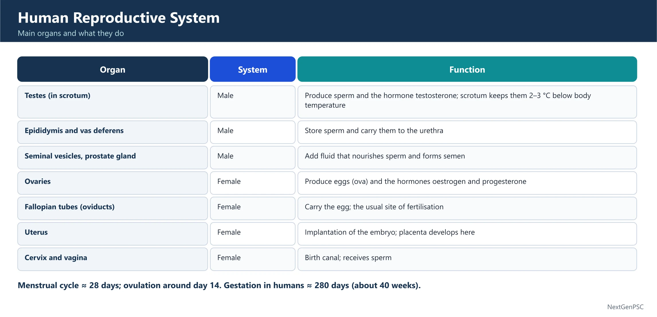
1. Primary Sex Organs: The Testes
The testes are a pair of oval-shaped glands responsible for producing sperm and hormones.
Scrotum: The testes are situated outside the abdominal cavity in a pouch called the scrotum. This is crucial because it keeps the testes 2–2.5°C cooler than the internal body temperature, a requirement for Spermatogenesis.
Seminiferous Tubules: Located inside the testes, these are the actual sites where sperm are produced.
Leydig Cells: These cells produce androgens, primarily Testosterone.
2. Accessory Ducts and Transport
Once produced, sperm must mature and travel through a series of ducts.
Epididymis: A coiled tube where sperm are stored and gain motility (the ability to swim).
Vas Deferens: A long muscular tube that carries sperm from the epididymis toward the urethra.
Ejaculatory Duct: Formed by the union of the vas deferens and the duct of the seminal vesicle.
3. Accessory Glands and Semen Production
The survival of sperm depends on the secretions from three main glands, which together with sperm form Semen.
Seminal Vesicles: Produce a sugar-rich fluid (fructose) that provides energy to sperm.
Prostate Gland: Secretes an alkaline fluid that helps neutralize the acidity of the female reproductive tract.
Bulbourethral (Cowper's) Glands: Release a lubricating fluid that also neutralizes any acidic urine remaining in the urethra.
4. External Genitalia
Penis: The external intromittent organ made of special erectile tissue. It facilitates the transfer of semen into the female reproductive tract.
Glans Penis: The enlarged tip covered by a loose fold of skin called the Foreskin (prepuce).
| Component | Source | Function |
|---|---|---|
| Sperm Cells | Testes | Genetic contribution (Fertilization) |
| Fructose | Seminal Vesicles | Energy source for sperm motility |
| Alkaline Fluid | Prostate Gland | Neutralizes vaginal acidity |
| Mucus/Lubricant | Bulbourethral Glands | Lubrication and urethral cleaning |
Prelims tips
- Spermatogenesis: Occurs in the seminiferous tubules; regulated by FSH and LH.
- Rete Testis: The network of tubules within the testis that connects to the vasa efferentia.
- Urethra: In males, it serves as a common passage for both urine and semen (Urogenital duct).
- Temperature: 2–2.5°C lower than body temperature is vital for viable sperm production.
Mains points
- Reproductive Health: Issues like low sperm count (Oligospermia) are increasingly linked to environmental pollutants, heat exposure, and sedentary lifestyles.
- Oncology: Prostate cancer is one of the most common cancers in men globally, highlighting the importance of regular screening after age 50.
- Endocrine Regulation: The Hypothalamic-Pituitary-Gonadal (HPG) axis is the primary control mechanism for male fertility; any disruption here can lead to infertility.
- Evolutionary Biology: The external placement of testes in a scrotum is an evolutionary adaptation found in most mammals to optimize the cooling needed for sperm stability.
Did you know?
- A single sperm takes about 64 to 72 days to fully develop.
- The Prostate gland is about the size of a walnut in young men, but it can grow much larger as men age, often causing urinary issues.
- Sperm are the only cells in the human body designed to live and function outside the body they were created in.
The Female Reproductive System: Structure and Cycle
The Female Reproductive System is a highly specialized biological network designed for the production of female gametes (ova), facilitating fertilization, and supporting the development of a fetus during pregnancy. Unlike the male system, it operates on a rhythmic 28-day Menstrual Cycle and includes specialized organs like the Uterus and Fallopian Tubes.
1. Primary Sex Organs: The Ovaries
The ovaries are a pair of almond-shaped glands located in the lower abdomen.
Function: They produce the female gamete (ovum) and secrete essential steroid hormones like Estrogen and Progesterone.
Egg Storage: A female is born with a finite number of primary follicles, which mature and are released one by one during the reproductive years.
2. Accessory Ducts: The Pathway to Life
These structures are responsible for transporting the egg and providing a site for fertilization.
Fallopian Tubes (Oviducts): These extend from the periphery of each ovary to the uterus. The Ampulla region is the specific site where Fertilization typically occurs.
Uterus (Womb): A hollow, pear-shaped muscular organ. Its innermost layer, the Endometrium, undergoes cyclic changes every month and is where the embryo implants.
Cervix: The lower narrow part of the uterus that opens into the vagina.
3. External Genitalia and Mammary Glands
Vulva: Includes the labia majora, labia minora, and the clitoris (a tiny sensory structure).
Mammary Glands: Paired structures (breasts) containing glandular tissue and fat. They are functional in females to provide nourishment (milk) to the newborn.
| Hormone | Source | Primary Role |
|---|---|---|
| Estrogen | Ovarian Follicle | Development of secondary sexual characters |
| Progesterone | Corpus Luteum | Maintenance of pregnancy & uterine lining |
| LH (Luteinizing) | Pituitary Gland | Triggers Ovulation (release of egg) |
| FSH | Pituitary Gland | Stimulates growth of ovarian follicles |
Prelims tips
- Fertilization Site: Always remember it happens in the Ampulla of the Fallopian tube.
- Ovulation: Triggered by a surge in LH (Luteinizing Hormone) on approximately the 14th day of the cycle.
- Endometrium: The layer that sheds during menstruation and supports the embryo during pregnancy.
- Corpus Luteum: Formed after ovulation; it secretes Progesterone to maintain the pregnancy.
Mains points
- Maternal Healthcare: High Maternal Mortality Rate (MMR) in developing regions is often linked to a lack of awareness regarding reproductive anatomy and prenatal care.
- Ethical Dimensions: Issues such as IVF, egg freezing, and surrogacy highlight the intersection of reproductive biology with modern law and ethics.
- Social Impact: Understanding the menstrual cycle is vital for ending the social stigma and 'period poverty' that affects millions of women globally.
- Pathology: Conditions like PCOS (Polycystic Ovary Syndrome) and Endometriosis are on the rise due to lifestyle factors, impacting fertility and quality of life.
Did you know?
- The Ovum (egg cell) is the largest cell in the human body, visible even to the naked eye (roughly the size of a needle point).
- The Uterus is one of the most flexible muscles in the body; it can expand to over 500 times its original size during pregnancy.
- A female baby is born with nearly 1-2 million eggs, but by the time she reaches puberty, only about 300,000 remain.
Nutrition: Biomolecules, Vitamins, Minerals and Deficiency Diseases
A balanced diet supplies carbohydrates, proteins and fats for energy and growth, and vitamins and minerals in small amounts – their lack causes deficiency diseases.
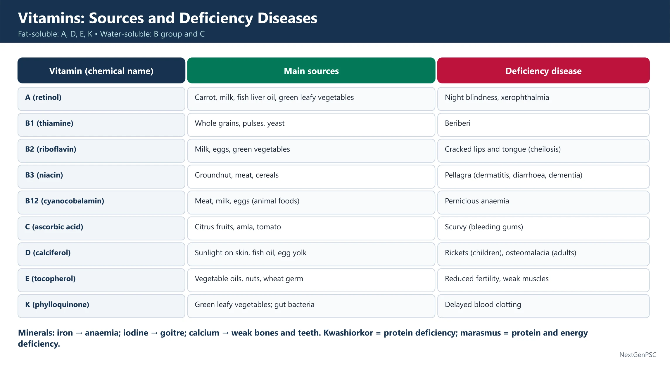
Macronutrients
Carbohydrates: main source of energy (about 4 kcal per gram) – sugars, starch, cellulose (roughage).
Proteins: body-building and repair (about 4 kcal per gram); made of amino acids. Deficiency causes kwashiorkor (protein lack – swollen belly) and marasmus (protein and calorie lack – wasting).
Fats: concentrated energy (about 9 kcal per gram); insulation and absorption of fat-soluble vitamins.
Vitamins
Fat-soluble (A, D, E, K): stored in the body. Water-soluble (B-complex, C): not stored; excess is excreted, so needed regularly.
Vitamin A (retinol): night blindness, xerophthalmia. Vitamin D (calciferol): rickets in children, osteomalacia in adults; made in skin by sunlight.
Vitamin E (tocopherol): antioxidant; deficiency linked to reproductive problems. Vitamin K: needed for blood clotting.
Vitamin B1 (thiamine): beriberi. B2 (riboflavin): cracked lips and tongue. B3 (niacin): pellagra. B9 (folic acid): anaemia, neural tube defects. B12 (cobalamin, contains cobalt): pernicious anaemia.
Vitamin C (ascorbic acid): scurvy (bleeding gums); found in citrus fruits and amla.
Minerals
Iron: haemoglobin – deficiency causes anaemia. Iodine: thyroid hormone – deficiency causes goitre. Calcium and phosphorus: bones and teeth. Fluoride: teeth (excess causes fluorosis). Sodium and potassium: nerve and muscle function.
| Vitamin | Chemical name | Deficiency disease |
|---|---|---|
| A | Retinol | Night blindness, xerophthalmia |
| B1 | Thiamine | Beriberi |
| B3 | Niacin | Pellagra |
| B12 | Cobalamin | Pernicious anaemia |
| C | Ascorbic acid | Scurvy |
| D | Calciferol | Rickets, osteomalacia |
| K | Phylloquinone | Poor blood clotting |
Prelims tips
- Fat-soluble vitamins: A, D, E, K; water-soluble: B-complex and C.
- Vitamin B12 contains cobalt; vitamin D is made in the skin by sunlight.
- Kwashiorkor = protein deficiency; marasmus = protein and calorie deficiency.
- Iodine deficiency – goitre; iron deficiency – anaemia; excess fluoride – fluorosis.
- Fats give about 9 kcal per gram; carbohydrates and proteins about 4 kcal per gram.
Mains points
- India faces a triple burden of malnutrition – undernutrition (stunting, wasting), hidden hunger (micronutrient deficiency) and rising obesity.
- Food fortification (iodised salt, fortified rice, double-fortified salt) and supplementation (IFA, vitamin A) address hidden hunger cost-effectively.
- POSHAN Abhiyaan, the mid-day meal (PM POSHAN) and the PDS link nutrition to education and food security.
- Dietary diversity – millets, pulses, fruits and vegetables – matters as much as calories.
Did you know?
- Sailors on long voyages once died of scurvy until citrus fruits were added to their rations.
- Vitamin B12 is the only vitamin containing a metal (cobalt).
- Amla is one of the richest natural sources of vitamin C.
Immunity, Skeleton, Muscles, Skin and Sense Organs
The immune system defends the body through innate and acquired immunity; the skeleton and muscles give support and movement; the skin and sense organs connect us with the environment.
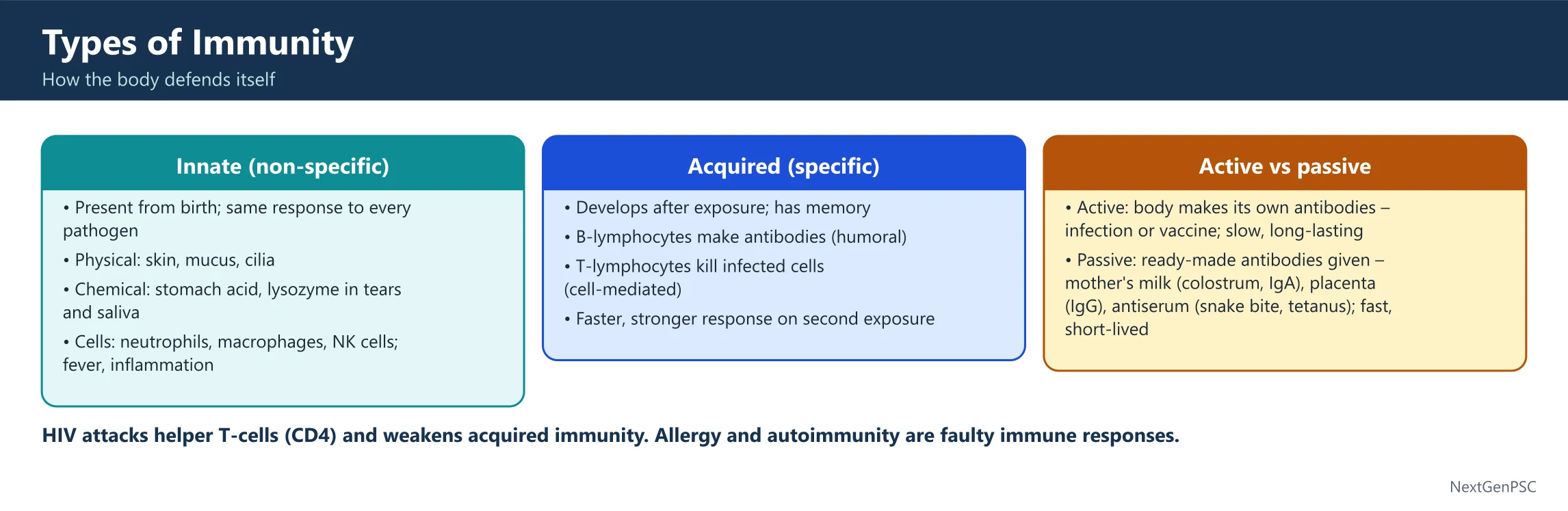
Immunity
Innate immunity: present from birth – skin, mucus, stomach acid, phagocytes (neutrophils, macrophages), fever and inflammation.
Acquired (adaptive) immunity: specific and has memory – B cells make antibodies (humoral immunity); T cells kill infected cells (cell-mediated immunity).
Active vs passive: active immunity follows infection or vaccination and lasts long; passive immunity comes from ready-made antibodies – mother's colostrum (IgA), antibodies crossing the placenta (IgG), antiserum (e.g. anti-venom).
Antibodies: IgG (most abundant, crosses the placenta), IgA (in milk and secretions), IgM (first to appear), IgE (allergy). HIV destroys helper T cells (CD4).
Skeleton and Muscles
Skeleton: an adult has 206 bones; the femur is the longest bone and the stapes (ear) the smallest. Ligaments join bone to bone; tendons join muscle to bone.
Muscles: skeletal (voluntary, striated), smooth (involuntary – gut, blood vessels) and cardiac (involuntary, striated, never tires).
Skin
Skin: the largest organ – epidermis (melanin gives colour), dermis (sweat glands, hair follicles); it protects, controls temperature and makes vitamin D.
Sense Organs
Eye: cornea, iris, pupil, lens and retina – rods for dim light, cones for colour; the blind spot has no receptors.
Eye defects: myopia (short-sight) – concave lens; hypermetropia (long-sight) – convex lens; presbyopia – bifocal lens; astigmatism – cylindrical lens; cataract – clouded lens.
Ear: outer ear, middle ear (malleus, incus, stapes) and inner ear – the cochlea for hearing and the semicircular canals for balance.
| Defect | Problem | Correction |
|---|---|---|
| Myopia | Cannot see distant objects clearly | Concave lens |
| Hypermetropia | Cannot see nearby objects clearly | Convex lens |
| Presbyopia | Age-related loss of near focus | Bifocal lens |
| Astigmatism | Uneven curvature of the cornea | Cylindrical lens |
Prelims tips
- B cells – antibodies (humoral); T cells – cell-mediated immunity; HIV attacks CD4 helper T cells.
- IgG crosses the placenta; IgA is in colostrum; IgE causes allergy.
- 206 bones in an adult; femur longest, stapes smallest.
- Rods – dim light; cones – colour vision.
- Myopia – concave lens; hypermetropia – convex lens.
Mains points
- Vaccination builds active immunity and herd immunity; Mission Indradhanush and the Universal Immunisation Programme aim at full coverage.
- Antimicrobial resistance weakens our ability to treat infections and is a major public health threat.
- Rising myopia among children is linked to screen time and less outdoor activity.
- Strong primary health care – screening, nutrition and immunisation – prevents many disorders of these systems.
Did you know?
- A newborn baby has about 300 bones; many fuse as it grows.
- The stapes in the middle ear is about 3 mm long.
- The skin of an adult weighs about 4 kg.